KNEE

Source: Wikipedia
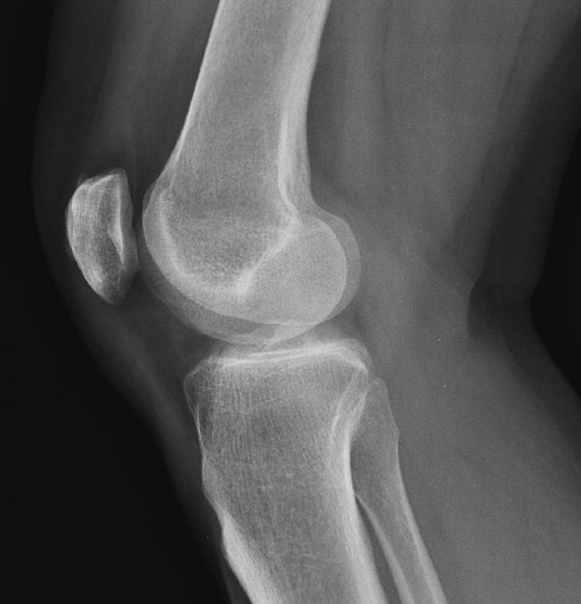
A knee joint effusion is usually radiographically appreciable as expansion of the suprapatellar bursa. This bursa usually contains a small amount of fluid and can often be seen as a thin, slightly radiopaque, finger-like projection situated posterior to the patella, separating the prefemoral and quadriceps fat pads.
Source: Radiopaedia
In about 84% of people, the suprapatellar bursa is contiguous with the joint itself, however in the remainder of the population, there is a full septum between bursa and the joint. That said, given that the joint capsule extends proximally to the patella, even the minority of patients with full septation may have a radiographically appreciable effusion which pushes the patella anteriorly and bulks out the suprapatellar space with radiodense fluid.
Knee joint effusions can be difficult to appreciate, particularly on patients with very fat or very muscular knees. With this in mind, care should be made when selecting exposure for bigger patients.
PRACTICAL POINT: when positioning for a lateral knee, try not to flex the knee much past 45°. There are a couple of reasons for this –
Firstly, overflexion may mask the presence of smaller joint effusions, as the patella is drawn into the trochlea and the suprapatellar space is compressed.
Secondly, we sometimes need to take measurements to diagnose patella alta/baja (patella too high or too low). The studies which figured out the normal ranges of ratio between patellar tendon length and patellar articular surface length (Caton-Deschamps index and Insall-Salvati ratio are the most commonly encountered) used specific degrees of flexion, therefore making measurements outside of these parameters reduces the validity of applying their normal ranges to measurements made.
Of course, some patients are just awkward and you have to take what you can get...
Trauma
A haemarthrosis refers to a joint effusion consisting of blood. It looks radiographically identical to any other flavour of joint effusion, however concurrent bony injury is a bit of a giveaway.
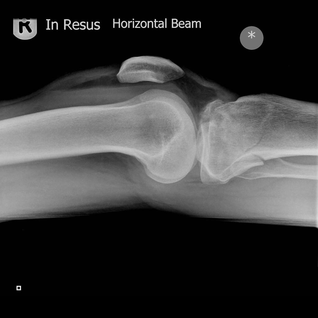
A lipoheamarthrosis describes layering of less radiodense fat on top of the more radiodense blood, in the same way that less dense oil will float on top of more dense water. This layering is appreciable on horizontal beam lateral projections as a discrete layer-line within the effusion.
Source: Radiopaedia
On occasion, a lipohaemarthrosis can appear as multiple layers, with a somewhat stripy appearance if the patient’s knee has not been supine for enough time for the components to separate fully, usually taking a couple of minutes or so.
The presence of a lipohaemarthrosis is pathognomonic for intra-articular bony injury; if no bony injury can be seen, further plain film views or a cross-sectional study may be required to diagnose the occult injury and inform management.
PRACTICAL POINT: when sending your HBL knee to PACS, ensure that the image is orientated as above, with the anterior/superior aspects to the left/right. Some radiographers and reporters prefer to view HBLs orientated longitudinally – this is largely a hangover from the bad old days of making multiple exposures on one physical film to save money and, in the opinion of opinionated people who run websites, has no place in the modern world of computed/digital radiography.
To confidently exclude the presence of a lipohaemarthrosis, the reporter must be certain that the image was taken horizontal beam. This means annotating and sending images correctly orientated.
Osteoarthritis

Source: Radiopaedia
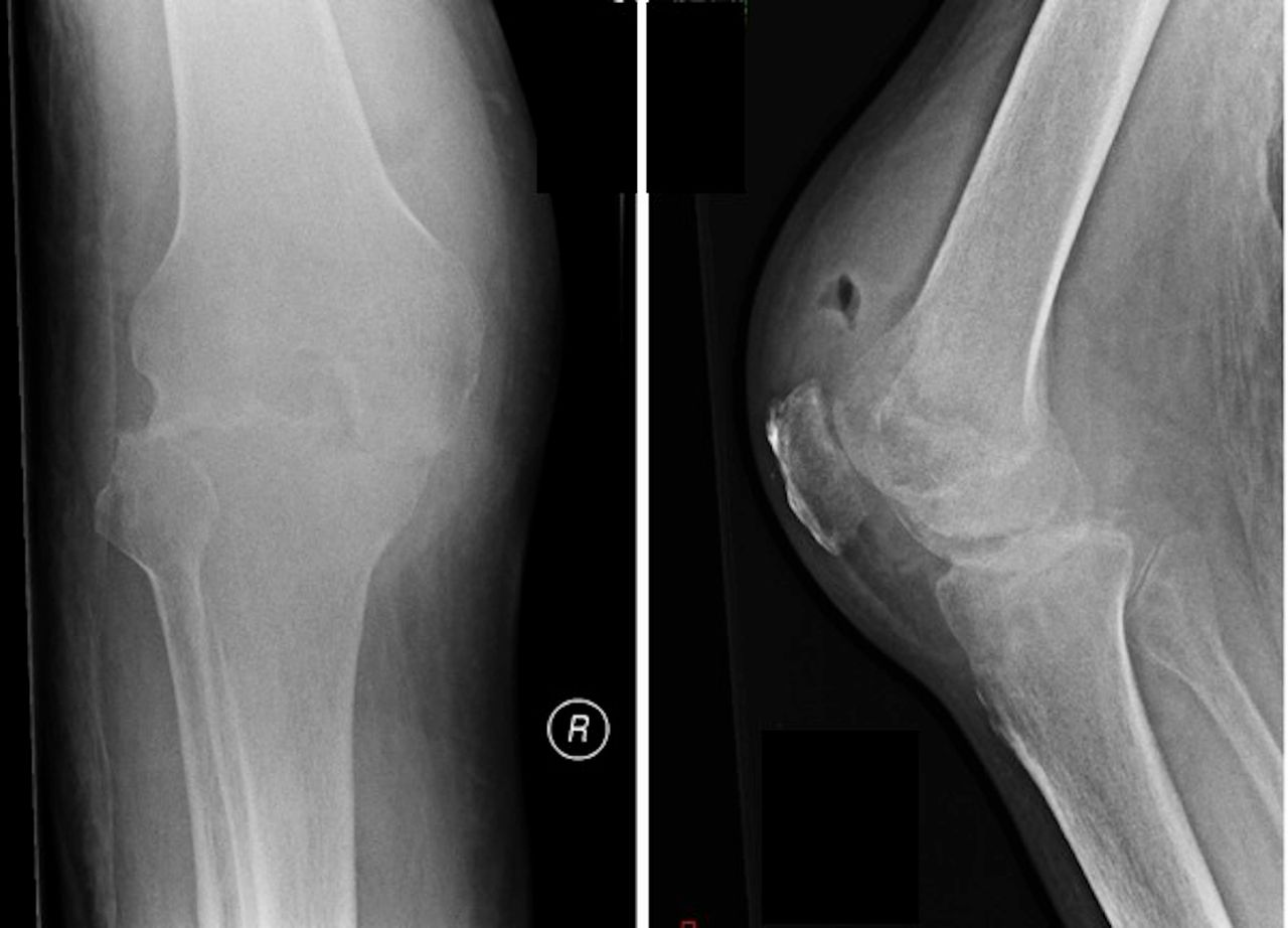
Osteoarthritis refers to degeneration of a joint due to repeated microtrauma. As the cumulative trauma outpaces the body’s ability to heal itself, radiographically appreciable changes occur. In the case above, there is a very large joint effusion, caused by the inflammatory response to insult as well as all four of the cardinal plain film radiographic signs of degenerative change (joint space narrowing, osteophytosis, articular sclerosis and subchondral cyst formation).
As a bonus, this patient also has chondrocalcinosis of their medial meniscus (often associated with but not necessarily indicative of OA; see CPPD).
Septic Arthritis
Source: BMJ Case Reports
Where osteoarthritis will cause joint space narrowing based on biomechanical pressures, generally resulting in one compartment being more affected than another, septic arthropathy will attack all articular surfaces within the affected joint simultaneously resulting in a more uniform distribution of joint space narrowing. Another feature is rapid progression, over weeks or months. Eventually, there will be destruction and collapse of the subchondral bone.
That said, septic arthropathy has quite specific clinical features and arthrocentesis is the proper objective test to make the diagnosis. Radiographs may demonstrate the effusion and the bony changes but, given that images may appear normal in the first couple of weeks after onset, negative imaging does not exclude septic arthropathy.
The role of imaging is to help exclude other potential causes of a red, hot, swollen joint, such as gout (look for tophi or mechanical erosions) or inflammatory arthropathy (look for juxta-articular osteopenia and erosions at the insertion of the joint capsule(bare area erosions)). Imaging findings (also involving MRI, for preference) would only form the basis of diagnosis if the joint is not able to be aspirated.