Effusions of the hip are infrequently seen and many authors consider them to be an absolute bugger to spot. This time we’re looking for the “Waldenstrom sign”.
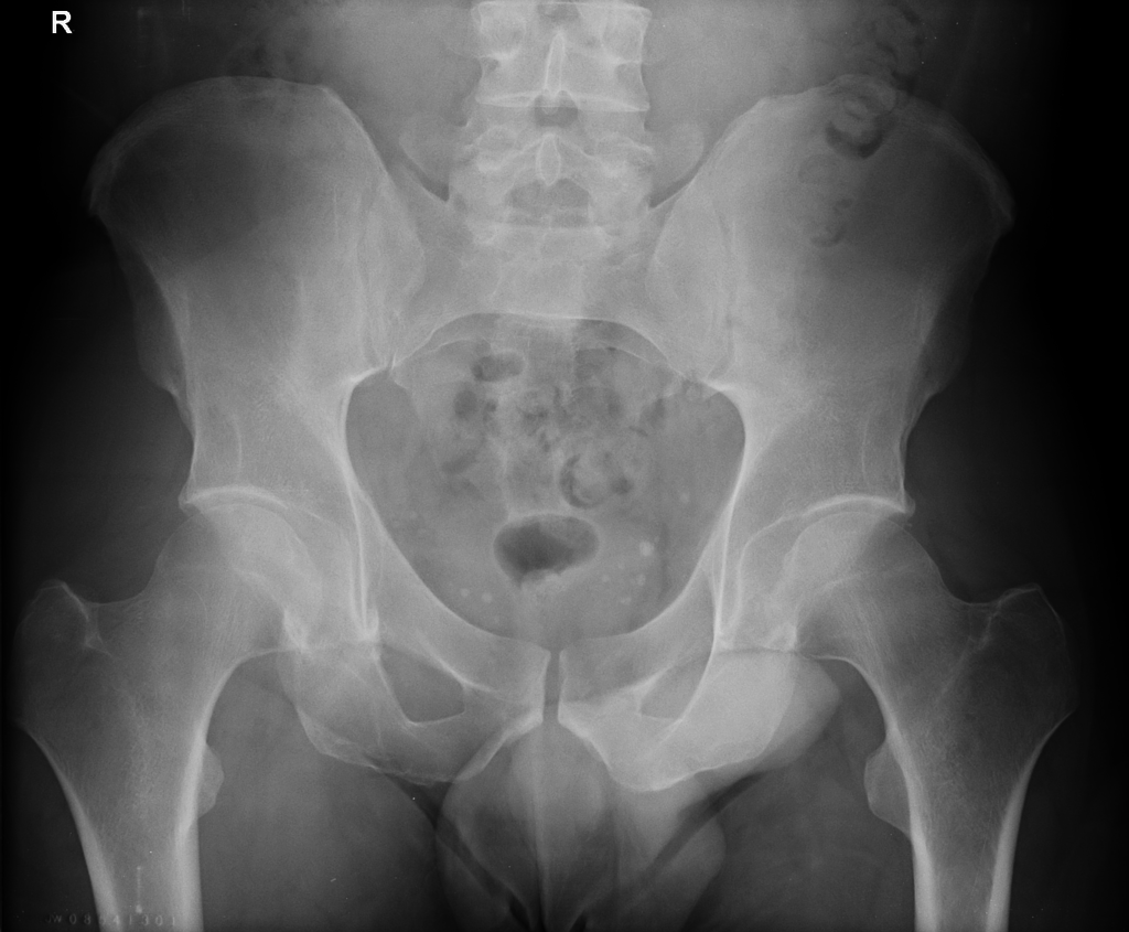
Consider the radiographic anatomy of the hip on a properly positioned, non-rotated AP pelvis -
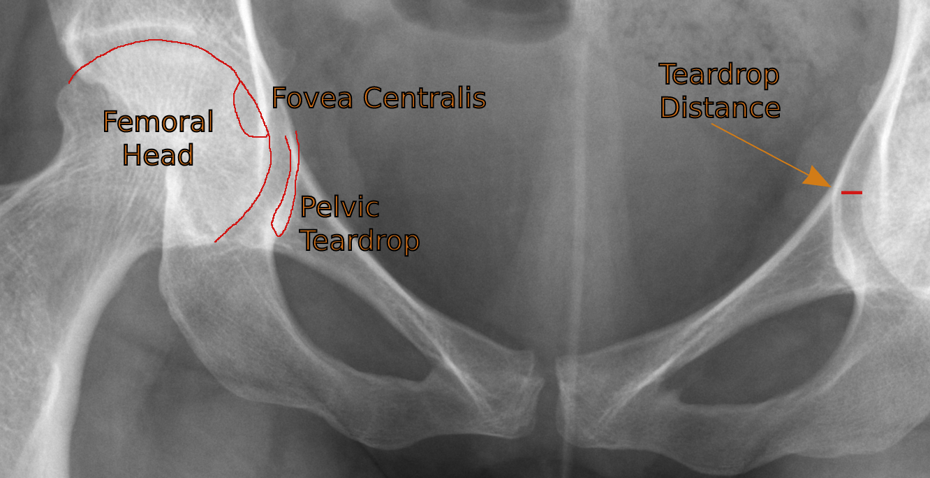
Normal hips (cropped)
After checking the obturator foramina for symmetry to judge whether or not the patient was rotated, we measure the teardrop distance from the most medial aspect of the femoral head to the adjacent lateral aspect of the pelvic teardrop (formed by the anteroinferior border of the acetabular fossa). A measurement of >11mm or >2mm compared to the contralateral side is considered positive for hip joint effusion.
Bear in mind the potential impact of chronic joint space narrowing when making these measurements and adjust interpretation accordingly.
Left sided effusion due to occult acetabular fracture
Source: Radiopaedia
While radiographic diagnosis of hip joint effusion is as non-specific as any other radiographic effusion diagnosis, aetiology can sometimes be determined by the clinical context. More common causes in adults are septic or inflammatory arthropathy.
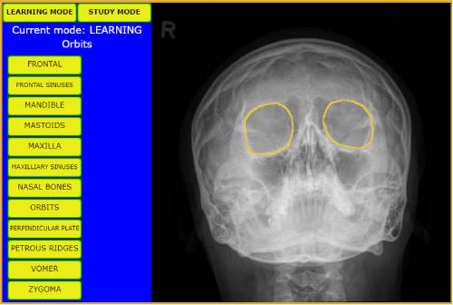
Learn bony anatomy from X-ray images - hover over the bones to display the names in learning mode, then test your knowledge and retention in study mode.
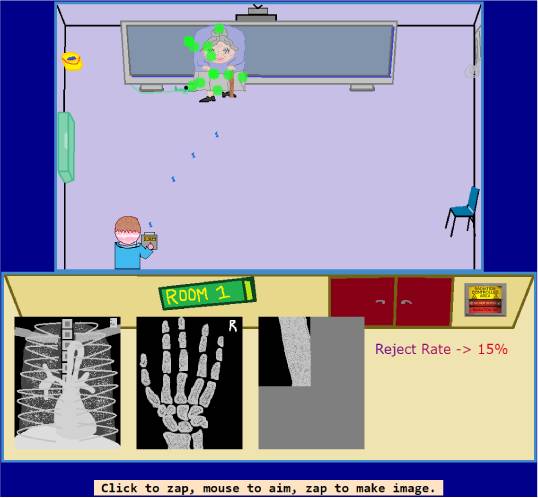
Radiology Quiz Machine - charge up the X-ray machine by filling the tank with photons generated by your expansive genius.